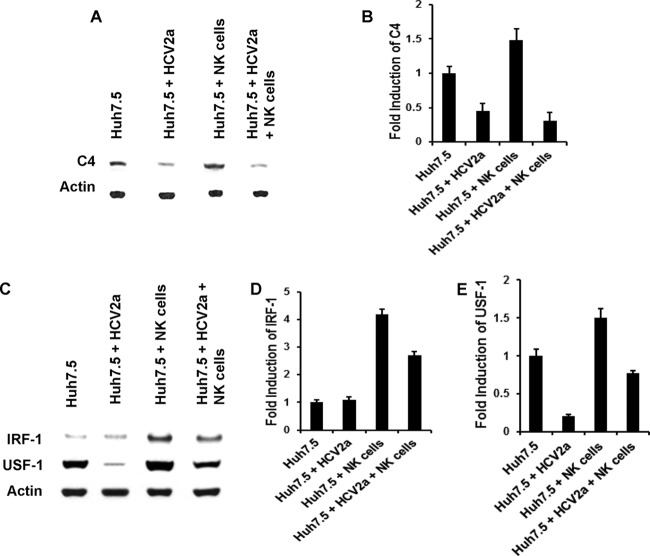
FIG 3.
NK cells fail to promote C4 augmentation in the presence of HCV. (A) rIL-2-activated NK cells were cocultured with Huh7.5 cells infected with HCV genotype 2a for 24 h. Mock-infected cells were cocultured similarly to the control. After removal of nonadherent NK cells, Huh7.5 cells were lysed and subjected to Western blot analysis for C4 protein expression. (B) Densitometric scanning of the Western blot displaying relative C4 inhibition. The results are from 3 independent experiments, with standard deviations indicated by error bars. The P value for the Huh7.5 control versus HCV 2a-infected cells was 0.0011, that for the control versus coculture with NK cells was 0.0179, and that for the control versus HCV 2a-infected cells and coculture with NK cells was 0.0006. (C) The expression status of transcription factors IRF-1 and USF-1 from cell lysates was examined by Western blotting. Cellular actin was used for comparison of protein loads in each experiment. (D and E) Results are from 3 independent experiments, with standard deviations indicated by error bars. The difference in IRF-1 expression between Huh7.5 control and HCV 2a-infected cells was not statistically significant (P > 0.05). However, changes in IRF-1 or USF-1 expression were statistically significant in comparison among Huh7.5 control and all other experimental samples (P = 0.004 to 0.0001).

