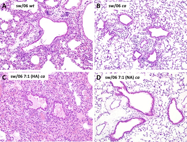
FIG 4.
H&E-stained sections of lungs from mice infected with sw/06 wt, sw/06 ca, sw/06 7:1(HA) ca, and sw/06 7:1(NA) ca viruses and sacrificed at 4 days postinfection. Lung pathology was most severe in animals infected with the sw/06 wt and sw/06 7:1(HA) ca viruses (A and C), whereas the lungs of mice infected with the sw/06 7:1(NA) ca virus (D) appeared relatively normal. Original magnification, ×200.

