FIG 3.

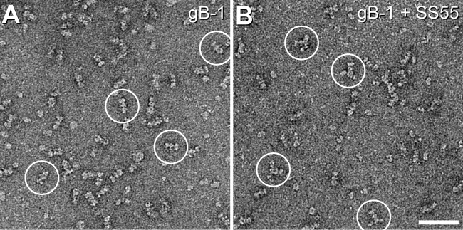
Negative-stain electron microscopy of gB. Shown are negatively stained low-magnification fields of gB1(730t) (A) and gB1(730t)-SS55 Fab (B). Some representative particles of each sample are circled. Bar, 50 nm.

Negative-stain electron microscopy of gB. Shown are negatively stained low-magnification fields of gB1(730t) (A) and gB1(730t)-SS55 Fab (B). Some representative particles of each sample are circled. Bar, 50 nm.