
FIG 5.
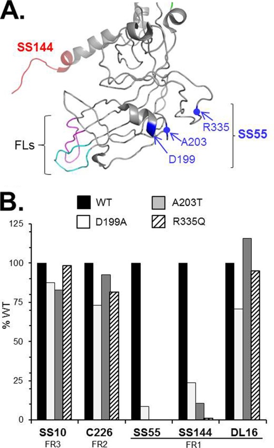
Identification of SS55 mar mutants. (A) Structure of FR1 from a gB monomer, based on the work of Stampfer et al. (6), with resolution of the loop between A328 and A338 at a low pH. The epitope of MAb SS144 is shown in red, and the fusion loops are shown in pink (FL1) and cyan (FL2). SS55 mar residues A203, R335, and D199 are highlighted in blue. Blue dots indicate the positions of A203 and R335 on the ribbon. (B) Full-length gB from transfected cell extracts was captured via PAb R242 (against the gB cytoplasmic tail) and was probed with anti-gB MAbs in an ELISA. Results of a representative experiment are shown. The percentage of WT absorbance was calculated as follows: (absorbance of test sample at 405 nm/absorbance of WT sample) × 100.