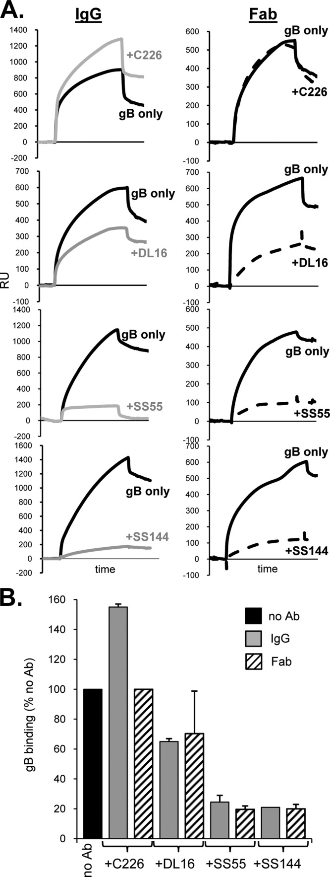
FIG 6.

Antibodies against neutralizing epitopes in FR1 block gB-liposome binding. (A) Fifty micrograms of each antibody (IgG or Fab) was incubated with 1 μg of purified, soluble gB(730t) for 1 h, and the complex was then flowed across liposomes bound to an L1 biosensor chip. Solid black curves represent the RU of gB binding to liposomes; dashed curves represent those for gB plus Fab (right); and gray curves represent those for gB plus IgG (left). The response from Fab or IgG flowing across liposomes only (no gB) was subtracted from the curves. (B) Bar graph representing the percentage of gB bound to liposomes for each sample. The percentage of gB bound was calculated based on maximal binding after 240 s as follows: (RU of test sample [after injection of gB-Ab]/RU of WT) × 100. Error bars indicate standard errors for at least two experiments.
