
FIG 4.
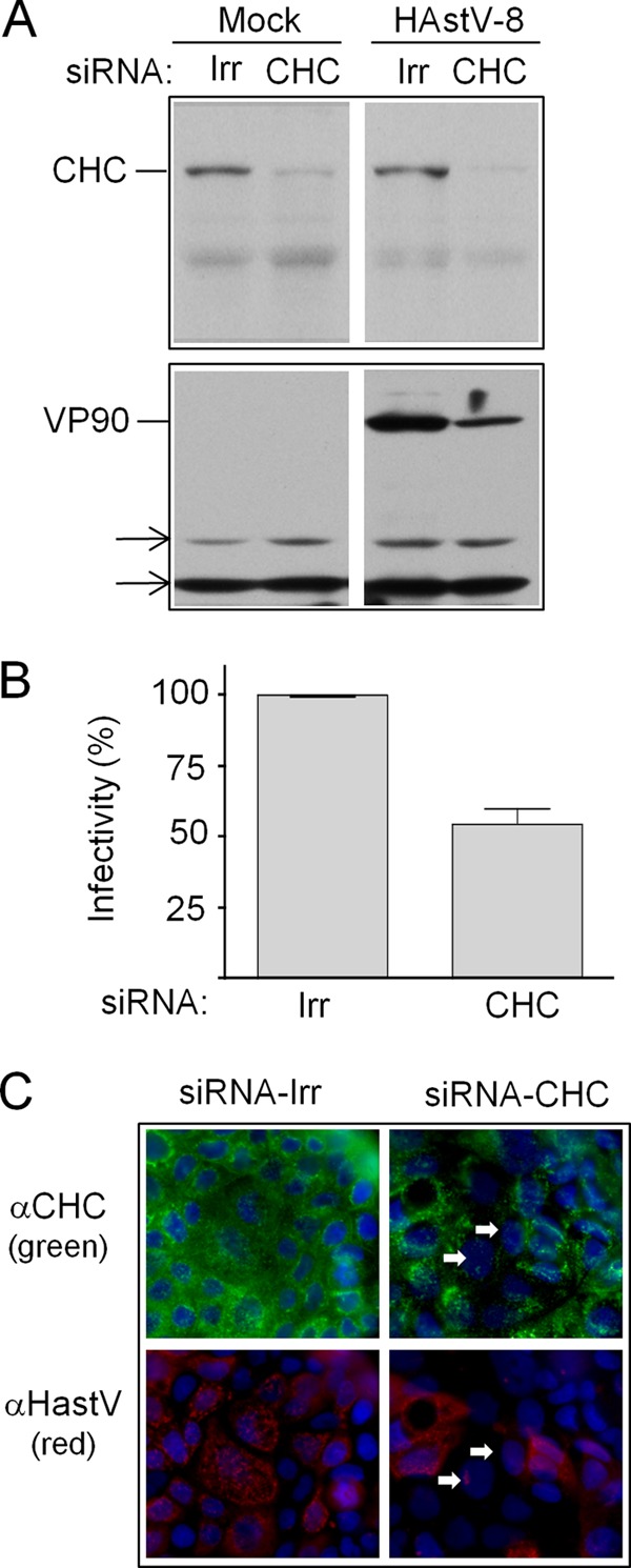
Effect of silencing the expression of CHC on HAstV-8 infectivity. (A) Caco-2 cells were transfected with the indicated siRNAs and, 72 h posttransfection, were infected with HAstV-8. Ten hours postinfection, the expression of CHC and VP90 was tested by Western blotting, using anti-HAstV-8 and anti-clathrin antibodies, as indicated. The arrows indicate cellular proteins that were nonspecifically recognized by the primary antibody and that served as loading controls. (B) Caco-2 cells were transfected with the indicated siRNAs and, 72 h posttransfection, were infected with HAstV-8. At 10 h p.i., the cells were processed for IF. Infectivity was determined by counting infected cells in cultures transfected with the indicated siRNAs. The data are expressed as percentages of the virus infectivity observed in cells treated with the irrelevant siRNA (Irr). The arithmetic means and standard deviations of three independent experiments performed in duplicate are shown. (C) Immunofluorescence assay showing the silencing of CHC. Caco-2 cells grown in coverslips were transfected with the indicated siRNAs, and 72 h posttransfection, the cells were infected with HAstV-8; at 10 h p.i., the cells were fixed and processed for IF using using anti-HAstV-8 (green) and anti-clathrin (red) antibodies, as indicated. Nuclei were stained with DAPI (blue). The arrows indicate cells transfected with the siRNA to CHC and infected with HAstV.