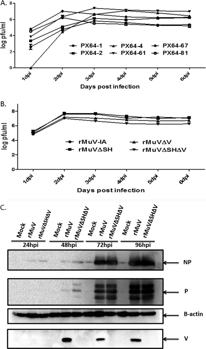
FIG 5.

Analysis of rMuVΔSHΔV in tissue culture cells. (A) Multicycle growth rate of rMuVΔSHΔV. Vero cells were mock infected or infected with rMuVΔSHΔV viruses from different rescues (PX64-1, PX64-2, PX64-4, PX64-61, or PX64-67) at an MOI of 0.1. Supernatants collected from culture medium of infected cells at 1, 2, 3, 4, 5, and 6 dpi were plated onto Vero cells for plaque assay, from which virus titer was calculated. (B) Multicycle growth curve of rMuVΔSHΔV virus compared to parent viruses. PX64-67 was selected for subsequent animal experiments. In vitro growth of rMuVΔSHΔV (PX64-67) was compared to rMuV, JL, rMuVΔV, and rMuVΔSH in Vero cells as described for panel A. (C) Viral protein expression levels of rMuVΔSHΔV at different time points postinfection. NP, P, and V protein expression levels in rMuVΔSHΔV-infected Vero cells (MOI of 0.01) were examined from 1 to 4 dpi and compared to those of rMuV infection.
