
Figure 2.
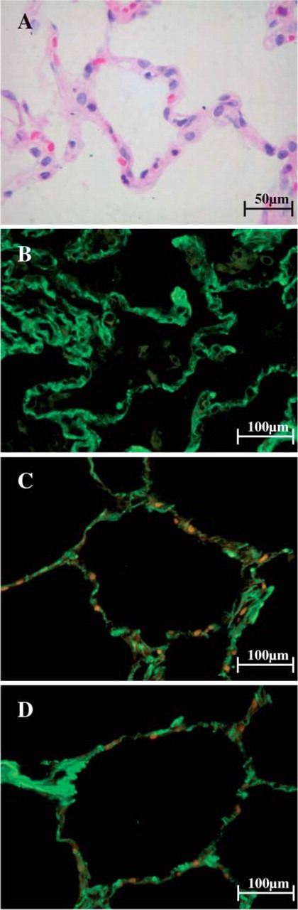
Morphological features of non-UIP lungs when stained with H&E and observed at optical microscope are characterized by preserved architecture and thin alveolar septa (A). After staining with fluorescein for types I, III, and V collagen fibers and observed at fluorescent microscope, alveolar walls of non-UIP lungs show fine green birefringence for type I collagen (B) and fine and minimal green birefringence for type III and type V collagen (C,D). Orange and red areas indicate the presence of elastic fibers and red cells, respectively.