Figure 1.
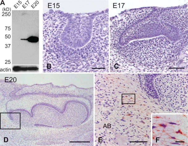
Western blotting analysis (A) and immunohistochemical staining of α-smooth muscle actin (αSMA) in the mandibular first molar at E15 (B), E17 (C), and E20 (D-F). Higher magnification of the boxed regions in D and E are shown in E and F, respectively. (A) α-SMA antibody reacted with cells in the E17 and E20 tooth germ. (B,C) α-SMA-positive cells are scarce in the tooth germs at the bud and cap stages. (D,E) The dental follicle cells around the cervical loop show α-SMA immunoreactivity at the early bell stage. (F) These cells exhibit long cell processes. AB, alveolar bone. Bars: B,E = 60 μm; C = 100 μm; D = 300 μm; F = 10 μm.

