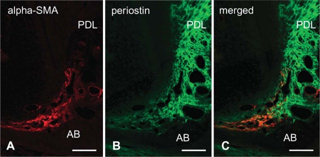
Figure 4.

Double-immunofluorescence staining of α-SMA (shown in red) and periostin (in green) in the apical root at P28. (A) α-SMA localization is seen in the dental follicle at the root apex. (B) Periostin is localized to the dental follicle and periodontal ligament (PDL). (C) The area of α-SMA immunopositivity exists on the alveolar bone (AB) side of the periostin-positive area. Bar = 100 μm.
