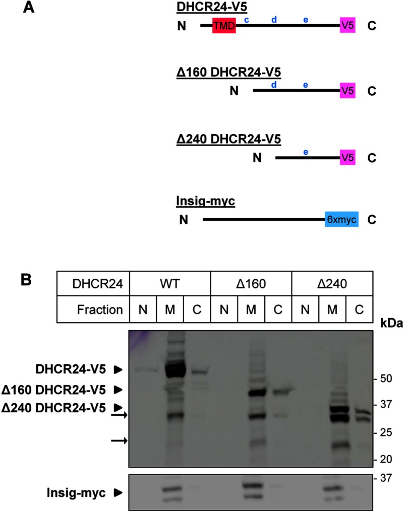
Figure 4. Membrane association of DHCR24 N-terminal truncations.
(A) The schematics of DHCR24-V5, Δ160 DHCR24-V5 and Δ240 DHCR24-V5 shown in relation to the putative TMD and other lower scoring putative TMDs (c, d, e) and Insig-myc. (B) CHO-7 cells were transfected with either 4 μg DHCR24-V5 (WT), Δ160 DHCR24-V5 (Δ160) or Δ240 DHCR24-V5 (Δ240), and co-transfected with 1 μg Insig-1-myc in a 10-cm dish for 24 h. Cell lysate was fractionated, and the nuclear (N), membrane (M) and cytoplasmic (C) fractions were separated by SDS–PAGE (10% gel) and immunoblotted with antibodies against V5 (DHCR24) and myc (Insig). Data from n=3 experiments.

