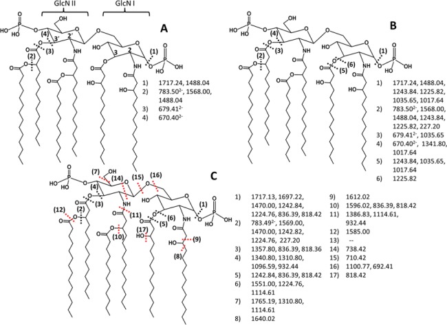
Figure 2.
MS/MS fragmentation maps of doubly deprotonated wild type E. coli lipid A (Mr = 1797.2) using (A) CID, (B) HCD, and (C) UVPD-MS. Each cleavage site is numbered, and the fragment ions arising from each cleavage site is listed. Those fragment ions that require multiple cleavages are listed next to each cleavage site. Red cleavages are only seen using UVPD-MS. The positions of the 2, 2′, 3, and 3′ carbons and the GlcN I and GlcNII are labeled in part A.

