
Figure 3.
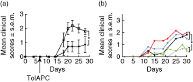
Effect of retinal antigen-pulsed tolerogenic antigen-presenting cells (TolAPC) on clinical course of experimental autoimmune uveitis (EAU) induced with interphotoreceptor retinoid-binding protein (IRBP). (a) Comparison of clinical scores of EAU mice treated with retinal extract-pulsed TolAPC (filled triangles, n = 7) or EAU mice not treated (open squares, n = 7). Data are shown as mean clinical EAU score ± standard error of the mean (s.e.m.) (ordinate) over time (abscissa). The retinal extract-pulsed TolAPC-treated mice show a significant (P ≤ 0·05) decrease in EAU clinical score over time compared to scores of EAU in untreated mice. (b) Antigen specificity of TolAPC-induced suppression. The EAU mice were treated with each type of TolAPC, 7 days post-induction of EAU (Materials and methods). Line graph of response of EAU mice to TolAPC pulsed with indicated antigens. The transferred antigen-presenting cells (APC) were not pulsed with antigen (black line, n = 15) or were treated with transforming growth factor (TGF)-β and pulsed with corneal extract (red line, n = 7), myelin basic protein (MBP) (blue line, n = 6), IRBP(1–20) (grey line, n = 14) or retinal extract (green line, n = 16). Data shown are mean clinical score (ordinate) over time (abscissa). An asterisk (*) indicates a significant difference between the areas under the curves. Statistics were performed using Prism software (Materials and methods).