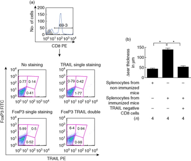
Figure 5.

Effect of CD8+ tumour necrosis factor (TNF)-related apoptosis-inducing ligand (TRAIL)– cells on suppression. (a) Flow cytometry gate used for analysis. Spleen cells were cultured with antigen-presenting cells (APC) treated with transforming growth factor (TGF)-β2 and interphotoreceptor retinoid-binding protein (IRBP). Seven days later the CD8+ T cells were enriched by magnetic beads and single-stained for CD8 (top panel). Lower panel: an aliquot of enriched CD8+ T cells was stained with TRAIL phycoerythrin (PE) (abscissa) and forkhead box protein 3 (FoxP3) (fluorescein isothiocyanate (FITC) (ordinate). (b) Local adoptive transfer assay using CD8+TRAIL– T cells. The experiment was performed twice with similar results. *Significant difference (P ≤ 0·05).
