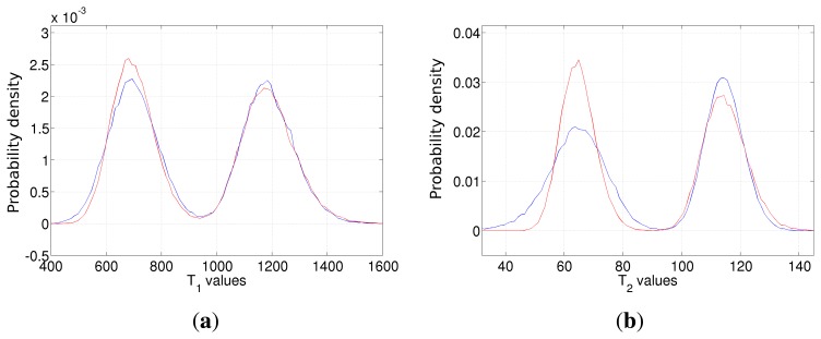
Figure 10.
The empirical probability density function of the T1 (a) and T2 (b) estimators in the case of Dataset 1 (blue line) and Dataset 2 (red line). Dataset 1 (blue line) imaging parameters are ideal for tissues with lower T1 and T2. On the contrary, Dataset 2 (red line) is ideal for the tissue with higher relaxation times.

