Figure 2.
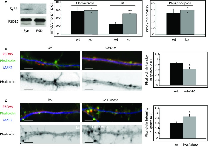
High SM levels accumulate in ASMko postsynaptic membranes and reduce the amount of filamentous actin.
A Western blots of the presynaptic and postsynaptic markers synaptophysin (Sy38) and PSD 95, respectively, in extracts containing the same amount of protein from total synaptosomal preparation (Syn) and from the postsynaptic enriched fraction (PSD). Graphs show mean ± s.d. of the levels of SM (P = 0.011) and cholesterol (in nmol/μmol phospholipids) and of phospholipids (nmol/mg protein) in postsynaptic membranes (PSD fraction) of wt and ASMko mice (n = 6).
B,C Top: Dendrites from cultured hippocampal neurons from wt mice treated or not with SM (B) or from ASMko mice treated or not with SMase (C) stained for MAP2 (blue), PSD95 (red), and phalloidin (green); bottom: phalloidin staining only. The graphs show mean ± s.d. of phalloidin fluorescence intensity per spine area (n = 250 dendritic spines from 3 independent cultures, *Pwt+SM = 0.02; *Pko+Smase = 0.03). Bars: 5 μm.

