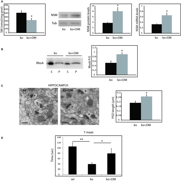
Figure 6.
- Mean ± s.d. of SM levels (nmol/mg protein) in synaptosomes from ASMko females treated or not with dexamethasone (n = 10; *P = 0.03). Western blot of NSM and tubulin levels in synaptosomes derived from ASMko females treated or not with dexamethasone. Graphs show mean ± s.d. of NSM protein (normalized to tubulin) and mRNA levels (n = 10, *PNSM prot = 0.024; *PNSM mRNA = 0.03).
- Western blots shows RhoA levels in supernatants (S) and pellets (P) after 100 000 g centrifugation of synaptosomes from ASMko females treated or not with dexamethasone. Graph shows mean ± s.d. of the RhoA ratio pellet/supernatant in synaptosomes from ASMko females treated or not with dexamethasone (n = 10, *P = 0.01).
- Electron micrographs of synapses in the hippocampal CA1 stratum radiatum of ASMko females treated or not with dexamethasone. Spines are indicated by asterisks. Graph shows mean ± s.d. of PSD length in μm (n = 70 synapses in each of 3 mice per condition, *P = 0.031).
- Results of the Y-maze test in wt, ASMko and dexamethasone ASMko treated females. Graph shows mean ± s.d. of the time (in seconds) spent by the mice in the novel arm (n = 7; **Pko vs wt = 0.009, *PDMko vs ko = 0.021).

