
Figure 1. Canalicular structure formation in control and LKB1 −/− mouse hepatocytes.
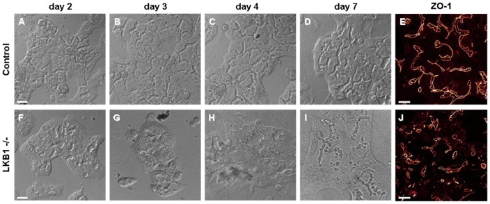
Progression of canalicular network formation was monitored in collagen sandwich cultures of mouse hepatocyte by DIC imaging. Elapsed times after cell isolation and seeding are indicated on the top. (A–D) In control cells, interconnecting canaliculi were gradually developed within 3–4 days. (E) Immunostaining of ZO-1 protein on day 6 suggests that canalicular structures are firmly sealed with tight junctions in these hepatocyte cultures (also see Video S1). (F–I) In contrast to control cells, LKB1 −/− hepatocytes developed only short and isolated canalicular structures. No interconnecting canaliculi were observed even 7 days after plating. (J) Similar result is obtained by anti-ZO-1 immunostaining performed on day 6. Note that despite the irregular morphology, tight junction proteins were present in the canaliculi of LKB1 −/− cells. Maximal projection of confocal images is shown. Scale bars 25 µm.