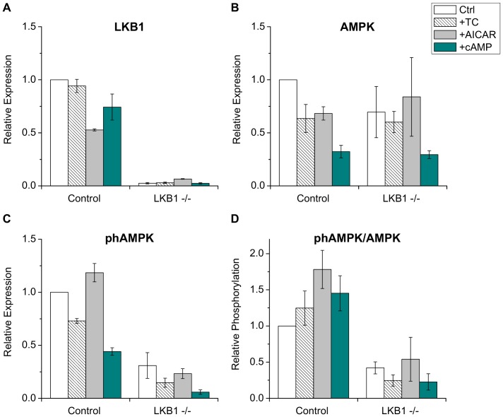
Figure 5. Expression and phosphorylation of AMPK.
Western blot analyses were performed with total cell lysates of cultured hepatocytes on day 6 using antibodies specific to LKB1 (A), AMPK (B), and phospho-Thr172 AMPK (phAMPK) (C). Immunoblots were quantified by densitometry; data were expressed as relative values to the expression level of untreated control cells. (D) Relative phosphorylation of AMPK was determined by dividing the phAMPK value by the total AMPK expression level. Pretreatments: TC – 100 µM taurocholate, AICAR - 500 µM AICAR, cAMP - 200 µM 8-Br-cAMP. Means ± S.E.M. of three experiments are shown. LKB1 expression is absent in the hepatocytes of the liver-specific LKB1 −/−, whereas AMPK expression is 64% of the control level. Both absolute and relative level of phosphorylated AMPK is greatly reduced in the LKB1-decifient hepatocytes.

