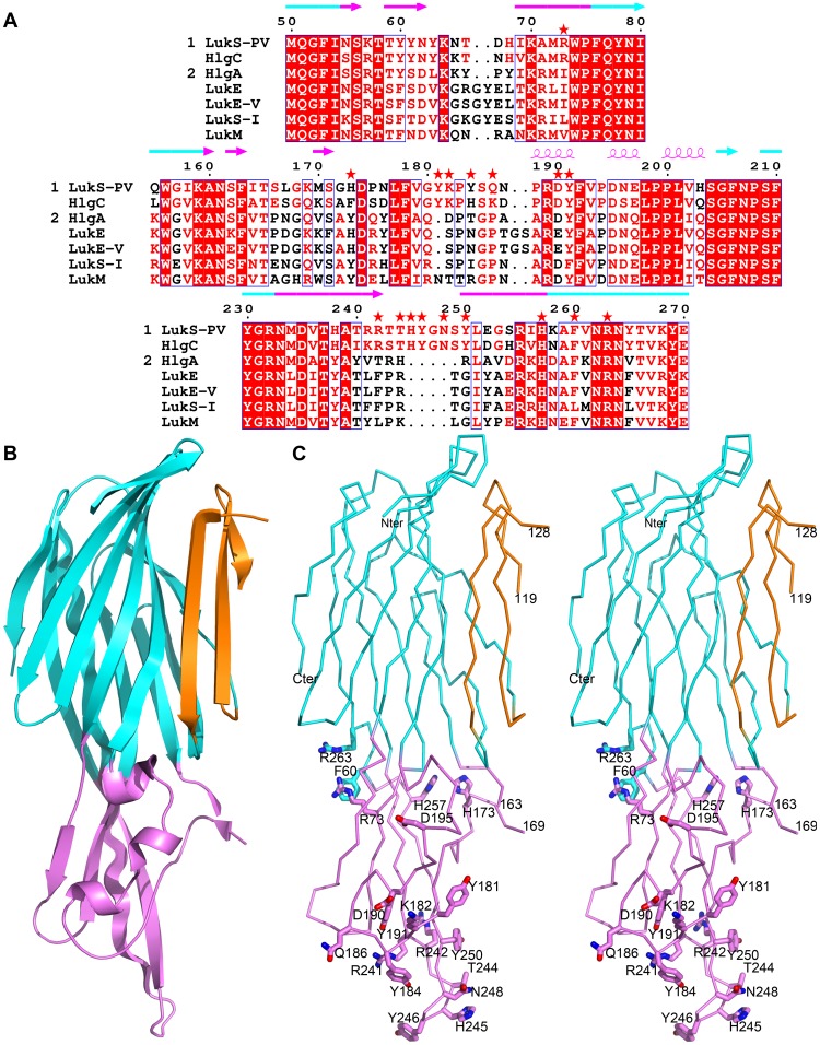
Figure 1. Positions selected for mutations in LukS-PV.
A. Sequence alignment of the three stretches of residues constituting the rim domain of class S components of leukotoxins. Numbering corresponds to the mature LukS-PV protein. Red asterisks indicate positions selected for mutation. Strictly conserved residues are indicated on a red background, while similar residues in group 1 (LukS-PV and HlgC) or in group 2 (all others) are indicated with red letters. The secondary structure of LukS-PV is indicated above the alignment, colored according to the corresponding structural domain: β-sandwich (cyan) and rim (purple). GenBank accession numbers are LukS-PV: CAA51251.1, HlgC: AAA26638.1, HlgA: AAA26637.1, LukE: CAA73667.1, LukE-V: BAB47174.1, LukS-I: CAA55782.1, LukM: BAA97866.1. B. Schematic representation of the three dimensional structure of LukS-PV [35], PDB entry 1T5R, highlighting the three structural domains: β-sandwich (cyan), stem (orange) and rim (purple). C. Stereo view of the Cα trace of wild-type LukS-PV. Residues selected for mutations are displayed as sticks and labeled.

