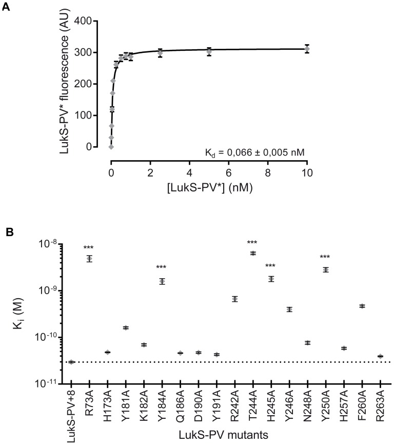
Figure 2. Binding properties of LukS-PV* and LukS-PV mutants to hPMNs and U937-C5aR cells.
A. Flow cytometry measurement of LukS-PV* and fluorescein-labeled LukS-PV G10C binding to human PMNs and U937-C5aR cells (n = 3). B. Graphic representation of the K i values obtained for wild-type or mutant LukS-PV. The dotted line corresponds to the value of wild-type LukS-PV. Error bars represent the 95% confidence interval. Statistical analysis: **: p<0.01, ***: p<0.001 (n = 3).

