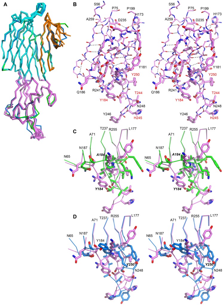
Figure 5. Structural variations of LukS-PV upon mutations in the rim domain.
A. Superposition of the Cα trace of all available structures of LukS-PV proteins, wild-type or mutants. The Y184A mutant structure is represented in green and the Y250A mutant structure in blue. For the other structures, the three structural domains are highlighted: β-sandwich (cyan), stem (orange) and rim (purple). B. Close-up stereo view of the rim domain of the LukS-PV wild-type structure, shown as stick (thin for main-chain and thick for side-chains). Residues labeled in red correspond to the most sensitive position as identified in this work. C. Stereo view of portion of the rim domain of wild-type LukS-PV (purple) and of the Y184A mutant structure (green). D. Stereo view of portion of the rim domain of wild-type LukS-PV (purple) and of the Y250A mutant structure (blue). Panels B to D, hydrogen bonds are represented as grey dots, the orientation of the image is the same as in A. Panels C and D, mutated positions are labeled in italic.

