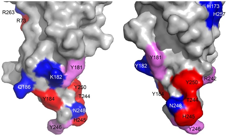
Figure 6. Molecular surface of the rim domain of LukS-PV.
Residues identified in this study as important for the binding of LukS-PV on the C5a receptor (Ki increased more than 50 fold upon mutation to Ala) are depicted in red. Mutated residues affecting binding to a lesser extent (i.e. increase in Ki by a factor between 5 and 50) are depicted in pink whereas residues for which no effect on binding was found upon mutation (increase in Ki less than 3 fold) are depicted in blue (Table 1). Two orthogonal views around a vertical axis are presented, with the orientation on the left being the same as in figure 5.

