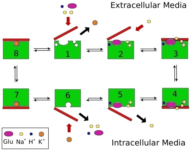
Figure 1. Cartoon showing the functional mechanism of the EAAT transporters.

Steps 3–4 denote the translocation of the binding site across the membrane with 3 Na+, H+, and glutamate bound to EAAT3, while steps 7–8 denote the same with only K+ bound. Step 2 shows the binding of the Na2 ion, which binds after the substrate and the closure of the HP2 gate. Step 5 shows the opposite happening in the intracellular media, assuming that the binding and unbinding of ligands are symmetrical.
