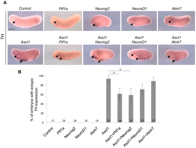
Figure 5. Comparison of the catecholaminergic inducing activities of five different bHLH factors.
(A) Whole mount in situ hybridization analysis of Tyrosine Hydroxylase (TH) expression on stage 24 embryos injected with the indicated mRNAs in one blastomere at the two-cell stage (anterior on the left, dorsal side up). Arrowheads indicate the position of previously described TH-positive antero-ventral neurons [53], while arrows point to ectopic TH staining. Note that, among all tested bHLH factors, only Ascl1 induces ectopic TH expression. Sibling embryos were also hybridized with the tubb2b probe as a positive control (see Figure 3A). (B) Quantification of embryos displaying ectopic TH+ neurons, showing that Neurog2 and Ptf1a significantly reduce the catecholaminergic inducing activity of Ascl1. Total number of analyzed embryos per condition is indicated in each bar. Error bars represent 95% confidence intervals. p<0,05 (*) (binomial test). Scale bar represents 500 μm.

