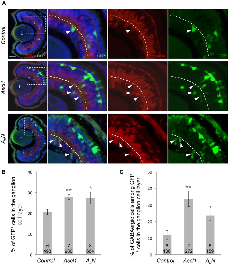
Figure 7. Retinal GABAergic inducing activity of Ascl1 resides in its basic domain.
(A) Stage 41 retinal sections immunostained with anti-GABA (red) and anti-GFP (green) antibodies. Embryos were lipofected with Ascl1 or the inducible ABN chimeric construct (encoding a Neurog2 protein with the basic domain of AsclI). Dexamethasone was added immediately after lipofection at stage 18. Panels on the right are higher magnifications of the dotted square delineated region. The dotted line indicates the inner plexiform layer. Arrowheads indicate GABA+/GFP+ cells within the ganglion cell layer. (B-C) Quantification of GFP+ cells and GABA+/GFP+ in the ganglion cell layer showing that similarly to Ascl1, ABN increases the percentage of cells in the ganglion cell layer but also biases these cells towards a GABAergic destiny. Total number of analyzed retinas and counted cells per condition is indicated in each bar. Values are given as mean +/– s.e.m. p<0,01 (**), p<0,05 (*) (Student’s t-test). L: Lens. Scale bar represents 50 μm.

