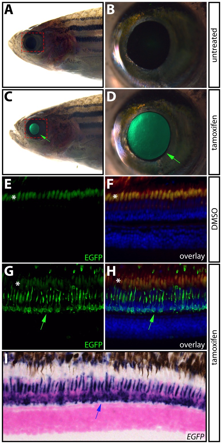
Figure 4. Inducible transgene expression in adult zebrafish.
(A–D) Expression of EGFP in the eye of a single adult Tg(krt5:Gal4-ERT-VP16; UAS:EGFP) zebrafish prior to (A and B) and after (C and D) treatment with 1 μM tamoxifen for one hour per day for three consecutive days. Green arrows point to EGFP expression in the eye. The areas bounded by the dashed red box in A and C are shown at high magnification in B and D, respectively. (E–H) Immunostaining of paraffin sections with anti-EGFP antibodies (shown in green) in eyes from DMSO- (E and F) and tamoxifen-treated (G and H) Tg(krt5:Gal4-ERT-VP16; UAS:EGFP) fish. Panels F and H show overlays with anti-EGFP antibody staining in green, Hoechst-stained nuclei in blue, and auto-fluorescence in red. Fish were drug treated as in A–D. Green arrows indicate EGFP expression in photoreceptors and asterisks (*) denote auto-fluorescence in photoreceptor outer segments. (I) In situ hybridization for EGFP mRNA in a paraffin section from a 4-OHT treated Tg(krt5:Gal4-ERT-VP16; UAS:EGFP) animal. The blue arrow shows EGFP expression in photoreceptors.

