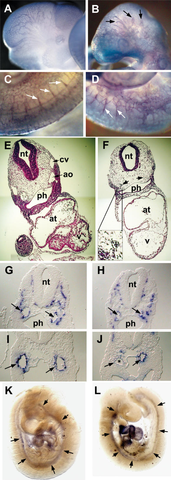
Figure 4.
Vascular defects in Hey1/2 DKO embryos. (A–D) Comparison of whole-mount PECAM antibody staining of E10.5 control (A,C) and DKO (B,D) embryos reveals intact vasculogenesis. Large cranial vessels appear truncated in mutants (arrows). Angiogenetic sprouts of intersomitic vessels (white arrows) are present, but the vascular pattern in the trunk is rather coarse. (E,F) H&E-stained cross-sections revealed reduction or loss of aorta (arrows) and cardinal vein. The myocardial wall is thinner in mutants, and ventricular trabeculation is missing. The neural tube is thinner and the mesenchymal compartment is cell-poor in mutants. (G,H) In situ hybridization with the endothelial marker VE-Cadherin identifies the aorta (arrows) and multiple smaller vessels. (I,J) The smooth muscle cell marker SM22 stains the aorta in controls (I), but in DKO embryos (J) staining is variable with partial or even complete loss (*) of the hybridization signal. (K,L) Whole-mount in situ hybridization for SM22 shows that the aorta is associated with smooth muscle cells along its length in controls and mutants, at least at this level of resolution. (ao) Aorta; (at) atrium; (cv) cardinal vein; (nt) neural tube; (ph) pharynx; (v) ventricle.

