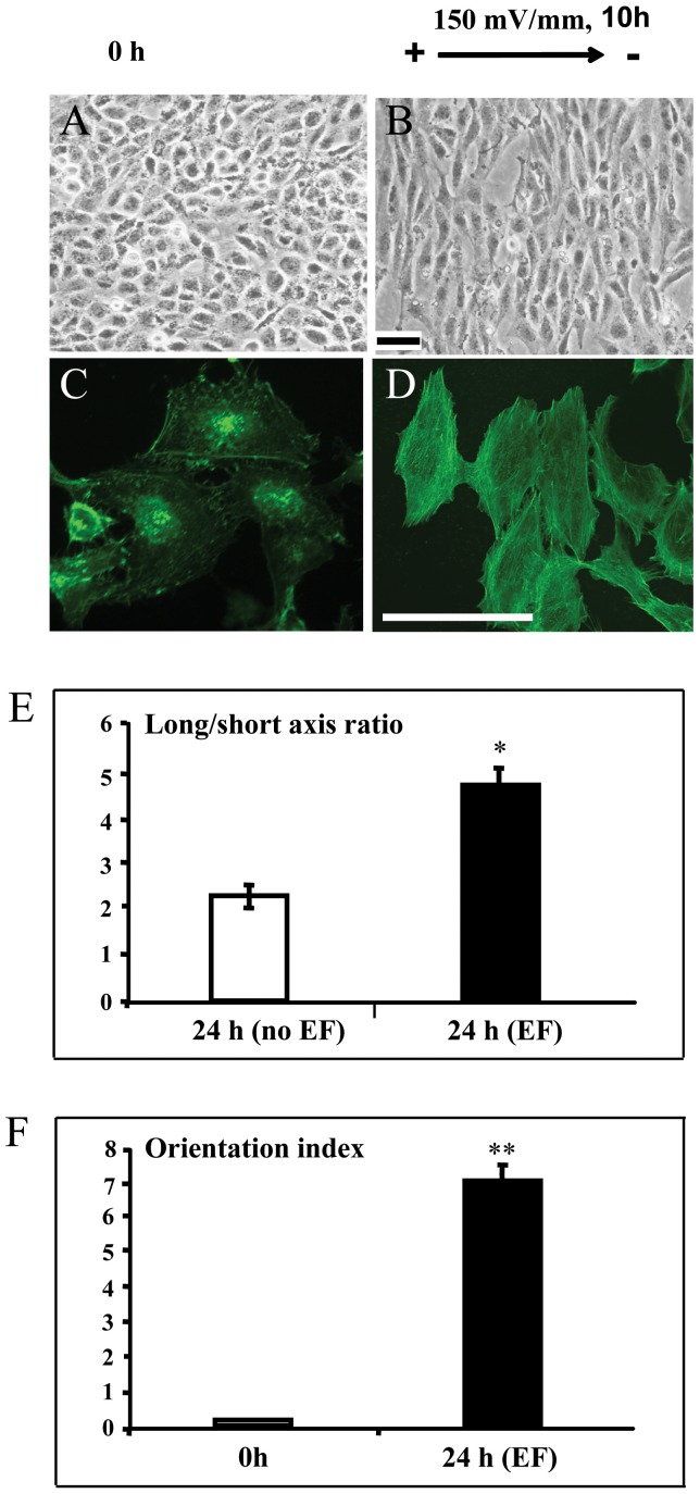
Figure 2. Trophoblast cells elongated and orientated perpendicularly in the electric field.
Trophoblast cells exposed to small, applied EF (150 mV/mm) showed elongation and perpendicular orientation (Fig 2B), while control cells that were not subjected to EF showed no such responsiveness (Fig 2A). The morphology of control and EF treated cells stained by fluorescein isothiocyanate labeled Phalloidin (for staining F actin) showed in Fig 2C and D, respectively. Enhanced elongation and orientation compared with non-EF stimulated cells (control) at field strength of 150 mV/mm (Fig 2E and F, respectively). The error bars represent the S.E.*p<0.05 and **p<0.0001. Bar = 50 μm

