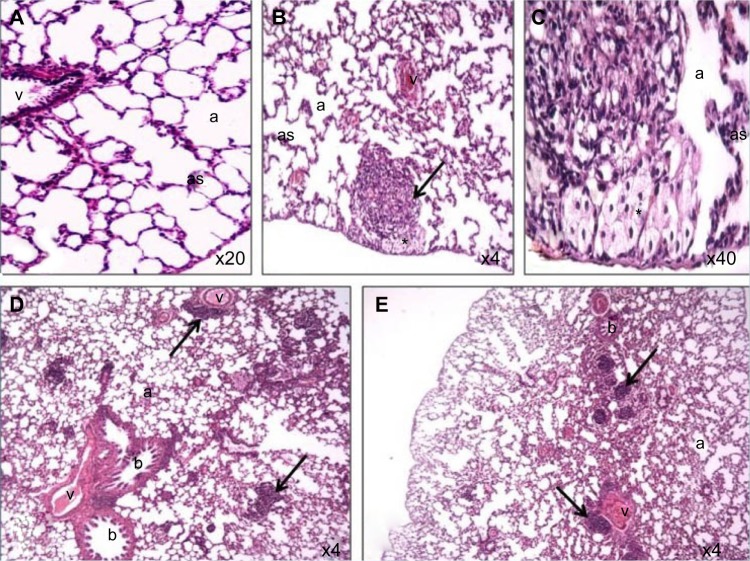
Figure 4.
Photomicrograph of lung tissue sections of rats treated with Ni nanoparticles.
Notes: (A) control (200X); (B) lymphocytic and eosinophilic infiltrates with thickening of alveolar walls and foamy macrophages (dark arrow) with the 20 mg/kg dose of Ni nanoparticles (40X); (C) foamy macrophages (indicated by the asterisk) with 20 mg/kg Ni nanoparticles (400X); (D) lymphocytic and eosinophilic infiltrates (dark arrow) with 1 mg/kg Ni nanoparticles (40X); (E) lymphocytic and eosinophilic infiltrates (dark arrow) with 10 mg/kg Ni nanoparticles (40X).
Abbreviations: a, alveolar space; as, alveolar septae; b, bronchioles; Ni, nickel; v, veins.

