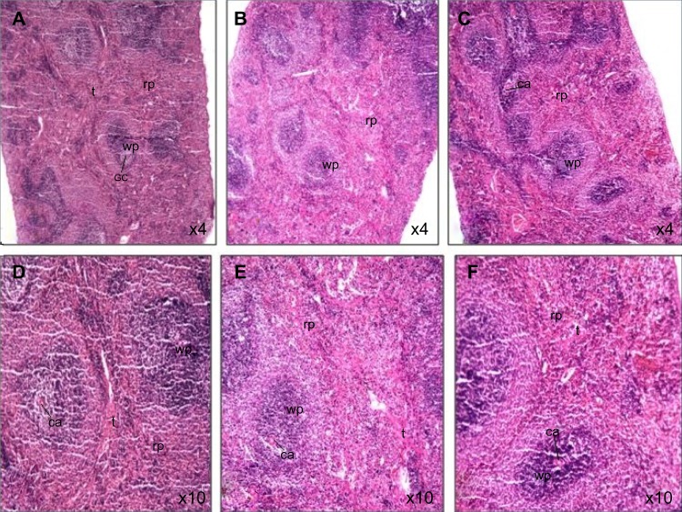
Figure 6.
Photomicrograph of spleen tissue sections of rats treated with Ni nanoparticles.
Notes: (A and D) Show normal spleen morphology with 1 mg/kg of Ni nanoparticles (X40 and X100, respectively); (B and E) show increased red pulp area with 10 mg/kg of Ni nanoparticles (X40 and X100, respectively); (C and F) show increased red pulp area with 20 mg/kg of Ni nanoparticles (X40 and X100, respectively).
Abbreviations: ca, central artery; gc, germinal center; Ni, nickel; rp, red pulp; t, trabeculae; wp, white pulp.

