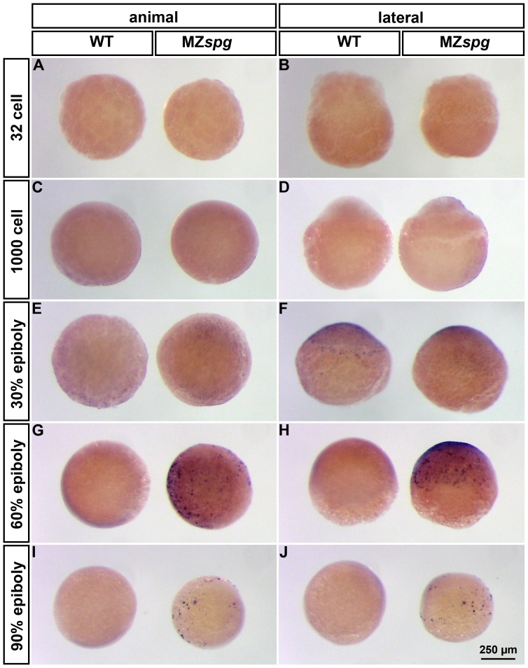
Figure 6. MZspg mutants have enhanced apoptosis during gastrulation.
TUNEL staining of WT (left embryo in each panel) and MZspg (right embryo in each panel) embryos at distinct embryonic stages between 32 cells and 90%-epiboly. Embryos are shown in animal (left column) and lateral (right column) view. Pou5f1 deficient embryos show enhanced apoptosis, starting at 60%-epiboly, compared to WT (G-J).

