Figure 3.
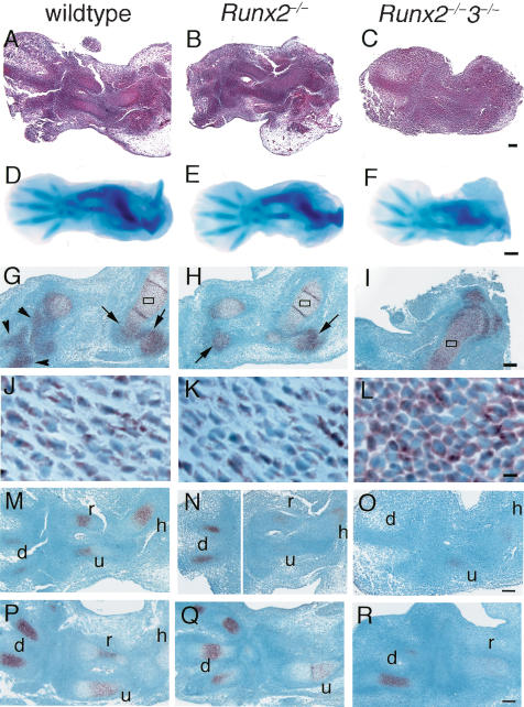
Early skeletal development in wild-type, Runx2–/–, and Runx2–/–3–/– embryos and in situ hybridization for Sox9 and Ihh expression. We used the forelimbs (A–C,G–R) and hind limbs (D–F) of wild-type (A,D,G,J,M,P), Runx2–/– (B,E,H,K,N,Q), and Runx2–/–3–/– (C,F,I,L,O,R) embryos for the analyses. (A–C) H&E staining at E12.5. (D–F) Whole-mount Alcian blue staining at E13.5. (G–L) In situ hybridization using Sox9 antisense probe at E13.5. (J–L) Higher magnification of boxed regions in G–I, respectively. (M–R) In situ hybridization using Ihh antisense probe at E12.5 (M–O) and E13.5 (P–R). We detected no signal using the sense probe of Sox9 and Ihh (data not shown). Cartilaginous anlagen was clearly seen in the wild-type (A) and Runx2–/– (B) embryos but not in the Runx2–/–3–/– embryos (C) at E12.5. Alcian blue staining of the hind limbs of E13.5 Runx2–/–3–/– embryos (F) was weak, compared with that of the wild-type (D) and Runx2–/– (E) embryos. (G,H,J,K) In the forelimb skeletons of wild-type and Runx2–/– embryos, the peripheral regions of the epiphyses of the humeri, radii, and ulnae (arrows in G,H), and most regions of the carpal and metacarpal bones (arrowheads in G) were composed of immature chondrocytes that strongly expressed Sox9, and the remaining regions of the humeri, radii, and ulnae, which were composed of differentiating chondrocytes, weakly expressed Sox9. (I,L) In the Runx2–/–3–/– forelimb skeletons, all of the chondrocytes strongly expressed Sox9. Ihh was strongly detected in immature chondrocytes. especially in digits of wild-type, Runx2–/–, and Runx2–/–3–/– mice. (h) humerus; (r) radius; (u) ulna; (d) digit. Bars: A–C,G–I,M–R, 100 μm; D–F, 500 μm; J–L, 10 μm.

