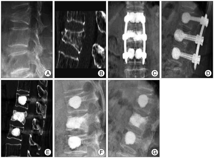
Fig. 3.
A case of a neurologically intact 72-year-old woman with a L1 burst fracture (Group B). A and B : Preoperative simple radiograph and computed tomography image showing about 60% height loss. C, D, and E : Simple radiographs and a computed tomography image taken at 12 months after percutaneous screw fixation showing consolidation of the fractured body and restored vertebral height. F and G : Simple flexion and extension radiographs taken at 13 months after screw removal reveal a well-maintained range of motion, which coincided with a completely pain free status.

