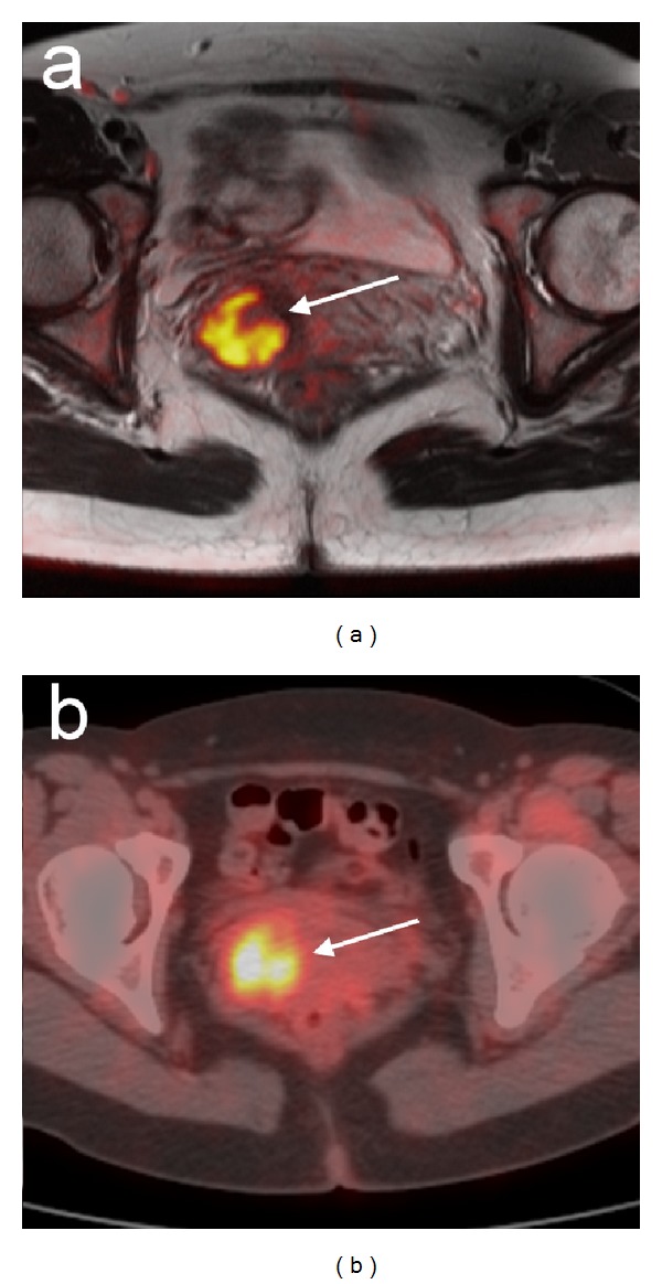
Figure 2.

An example of PET/CT and MRI in the female pelvis. A 43-year-old female patient with a primary well-differentiated adenocarcinoma of the uterine cervix. Primary cervical tumor is highlighted (arrow) and well correlated in (a) diffusion-weighted MRI and (b) 18F-FDG PET/CT. Reprinted with permission from [56]. Copyright 2008 Springer-Verlag.
