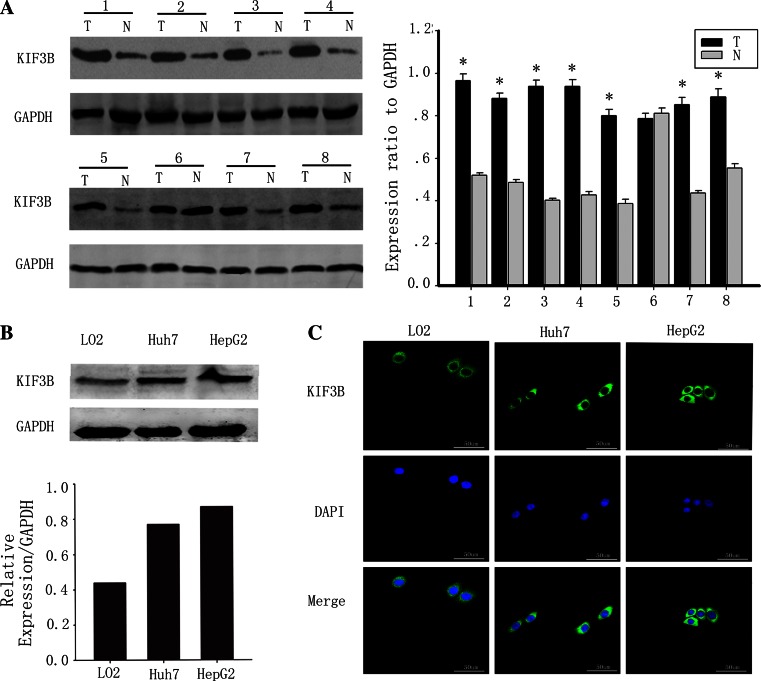
Fig. 1.
KIF3B is overexpressed in hepatocellular carcinoma but low or non existent in paired adjacent non-tumorous tissues. a Western blots of eight representative paired samples of hepatocellular carcinoma tissue (T) and adjacent non-tumorous tissues (N) immunoblotted against KIF3B. Whole-cell lysates were prepared from tissue specimens obtained from hepatocellular carcinoma and adjacent non-tumorous tissues. In all samples tested, KIF3B expression levels were significantly higher in hepatocellular carcinoma than in paired adjacent non-tumorous tissues. The bar chart demonstrates the ratio of KIF3B protein to GAPDH by densitometry. The data are mean ± SD of three independent experiments (*P < 0.01, tumor tissues compared with adjacent non-tumorous tissues). b The KIF3B protein expression was upregulated in the human hepatocellular carcinoma cell lines HepG2 and Huh7 cells, compared with the normal liver cell line LO2. c The KIF3B expression (green, mainly in the cytoplasm) in the LO2, Huh7 and HepG2 cells. The cell nuclei (blue) were stained with DAPI. Original magnification, ×400

