Figure 3.
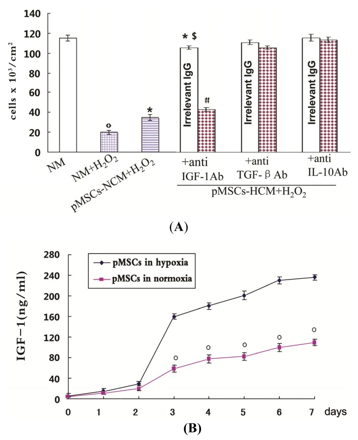
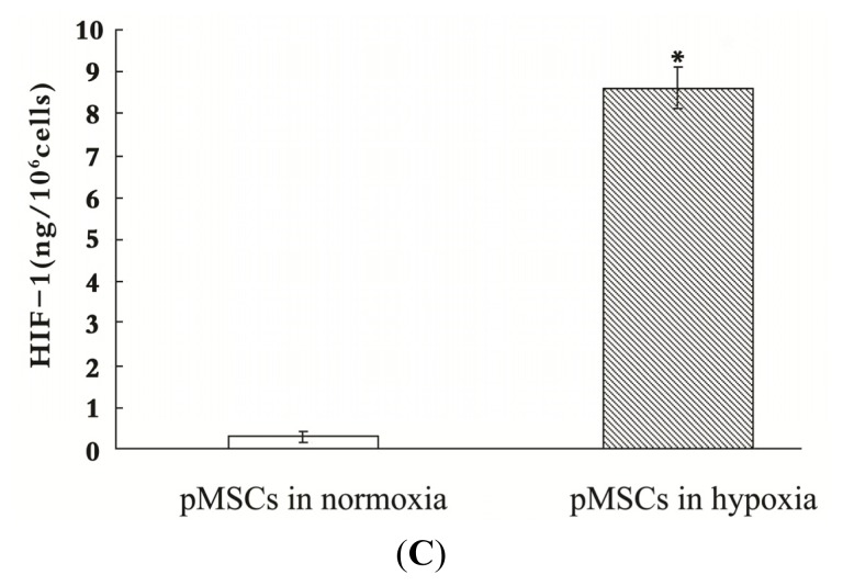
The pMSCs hypoxia culture mediums produce a better protective effect on caco2 cells from H2O2 through more IGF-1. (A) Effect of anti-IGF-1, anti-TGF-β and anti-IL-10 Ab on protective effect of pMSCs-HCM. Three hours after culturing H2O2-treated caco2 cells in pMSCs-HCM, cells were exposed to specific functional blocking Ab against IGF-1, TGFβ, L-10 or the corresponding irrelevant IgG. Cell count was evaluated after five days, º p < 0.05 versus NM; * p < 0.05 versus NM + H2O2; $ p < 0.05 versus pMSCs-NCM + H2O2; # p < 0.05 versus corresponding irrelevant IgG. (n = 6); (B) Hypoxia induced more IGF-1 secreted by pMSCs. The secretion of IGF-1 by pMSCs in both normoxia and hypoxia increased with time. There was no difference in the early two days. From the third day on, the secretion by pMSCs cultured in hypoxia was much higher than that in normoxia, and the difference persisted at seven days, º p < 0.05 versus pMSCs in hypoxia; and (C) Activated HIF-1 of pMSCs in hypoxia or normoxia measured by HIF-1 ELISA. pMSCs were cultured in hypoxia or normoxia for seven days and then the amount of activated HIF-1 was detected. Figure 3C shows mean amounts of activated HIF-1 normalized to amounts per million harvested cells. pMSCs in hypoxia have much more activated HIF-1 than that in normoxia; * p < 0.05 versus pMSCs in normoxia.


