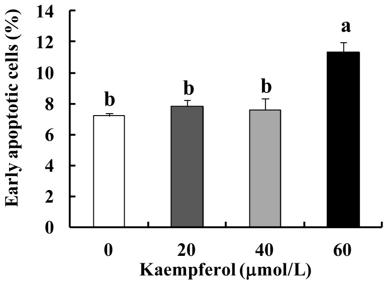
Figure 3.
Effect of kaempferol on apoptosis of SW480 cells. Cells were plated in 24-well plates and treated with various concentrations of kaempferol for 48 h. Cells were trypsinized, loaded with Annexin V and 7-AAD, and analyzed by flow cytometry. The percentages of early apoptotic cells were calculated. Each bar represents the mean ± SEM (n = 3). Means without the same letter (a or b) are significantly different (p < 0.05).

