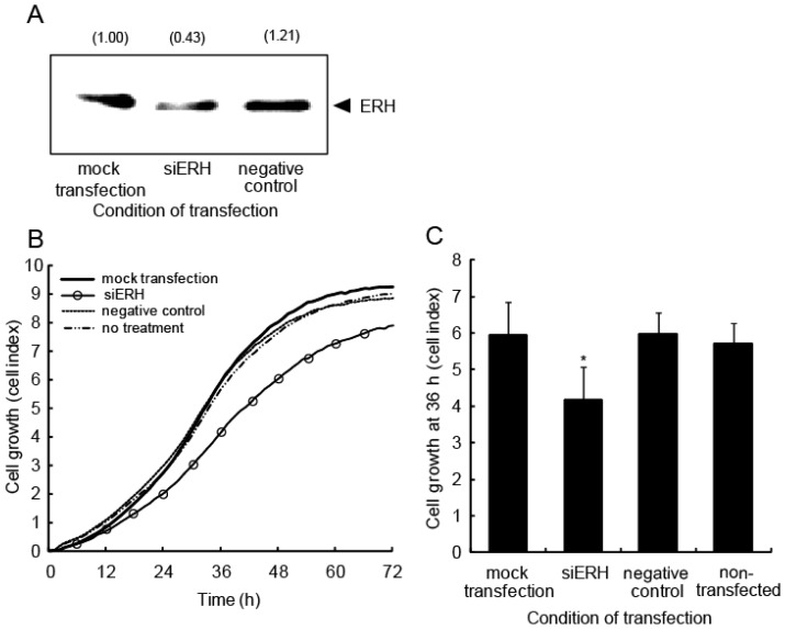
Figure 8.
Cell growth is delayed when ERH is knocked down in A549 cells. (A) A549 cells were transfected with siRNA oligonucleotides for either ERH or a negative control at 5 nM. Total cell extracts were prepared 48 h after transfection. The expression level of ERH was confirmed by western blot analysis. The relative expression level of ERH protein was quantified and is indicated in the panel; (B) A549 cells transfected with siRNA for ERH were incubated for 24 h and then re-plated into the wells of a device for RT-CES. Each experiment was conducted four times; (C) Cell growth was assessed using the cell index at 36 h. * p < 0.05 vs. mock-transfected cells.

