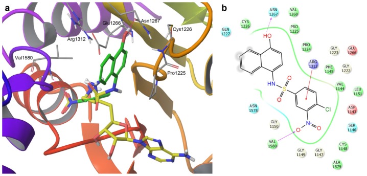
Figure 4.
(a) Induced-fit docking pose of SW155246 (carbon atoms in green) with human DNMT1. The co-crystal sinefungin (carbon atoms in yellow) is shown for reference; (b) Two-dimensional interaction diagram of the binding model of SW155246. Acidic, hydrophobic, basic, polar, and other residues at the active site are represented by red, green, purple, blue, and gray spheres, respectively. Hydrogen bonds between the ligand and backbone or side chains are shown in dashed pink lines. The π-cation interaction is shown with a red line.

