Fig. 1.
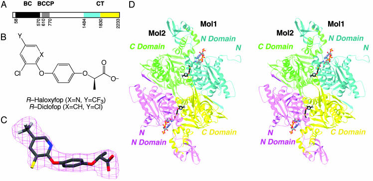
Crystal structure of CT domain in complex with haloxyfop. (A) Domain organization of yeast ACC. The N and C subdomains of CT are colored in cyan and yellow, respectively. (B) Chemical structures of the herbicides (R)-haloxyfop and (R)-diclofop. (C) Final 2Fo – Fc electron density at 2.8-Å resolution for haloxyfop, contoured at 1σ. (D) Schematic stereodrawing of the structure of yeast CT domain dimer in complex with haloxyfop. The N domains of the two monomers are colored in cyan and magenta, and the C domains are colored in yellow and green. The inhibitor is shown in stick models, in black for carbon atoms. The CoA molecule is shown for reference (11), in gray. C was produced with setor (28), and D was produced with ribbons (29).

