Figure 1.

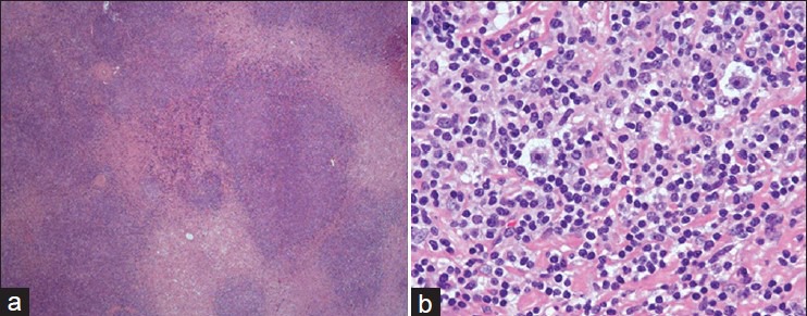
Photomicrograph of the lymph node biopsy (a) showing nodular sclerosis type of Hodgkin's lymphoma (H and E, ×20) and (b) high power view showing Reid–Sternberg cells (H and E, ×40)

Photomicrograph of the lymph node biopsy (a) showing nodular sclerosis type of Hodgkin's lymphoma (H and E, ×20) and (b) high power view showing Reid–Sternberg cells (H and E, ×40)