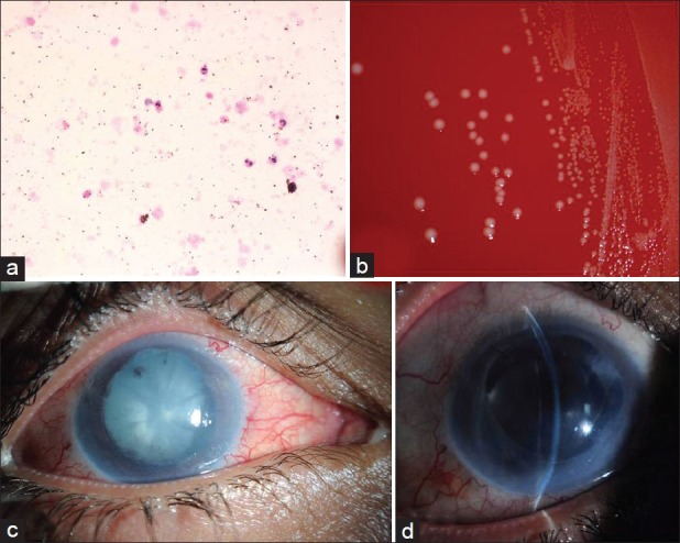
Figure 2.

(a) Histologic smear of the aqueous showing the predominant polymorphonuclear leukocytes (hematoxylin and eosin, original magnification ×400). (b) Blood agar plate showing the bacterial colonies with surrounding beta hemolysis. (c) The clinical appearance of the right eye cataract and posterior synechiae. (d) Postoperative slit-lamp photo of his right eye with intraocular lens (IOL) in the capsular bag
