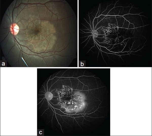
Figure 1.

(a) Colour fundus photograph showing macular serpiginous choroiditis with activity at temporal edge. (b) Fundus fluorescein angiogram (FFA) image showing distinct hyperfluorescence at the inactive nasal edge of the lesion. (c) Late FFA image showing fuzzy hyperfluorescence of active temporal edge, while hyperfluorescence at healed edge remain distinct
