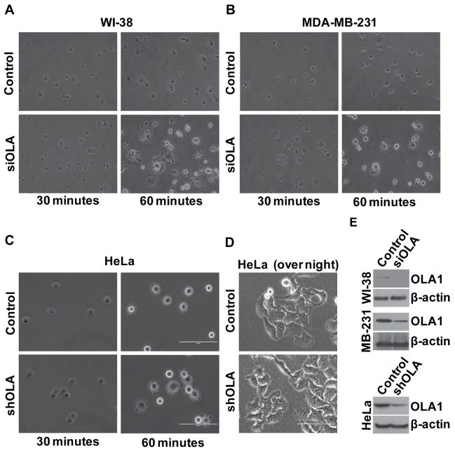
Figure 1. Down-regulation of OLA1 results in accelerated cell adhesion and spreading.
A–B, Human lung WI-38 cells (A) and breast cancer MDA-MB-231 cells (B) were transiently transfected with the control and OLA siRNA (siOLA), and 48 hours later the cells were plated on fibronectin-coated 6-well plates. Cell images were taken under an inverted light microscope at 30 and 60 minutes after plating. C, HeLa cells stably transfected with non-target shRNA (control), or OLA1-specific shRNA (shOLA) were plated on fibronectin-coated plates and imaged at 30 and 60 minutes after the plating. D, HeLa cells stably transfected with control or shOLA vectors were plated and imaged after an overnight incubation. All the above results represent at least three independent experiments. Scale bars: 100 μm. E, Cell lysates were immunoblotted to verify the effectiveness of OLA1-downregulation in the above knock-down experiments. The blot was re-probed with anti-β-actin antibody to check the equal loading of protein samples.

