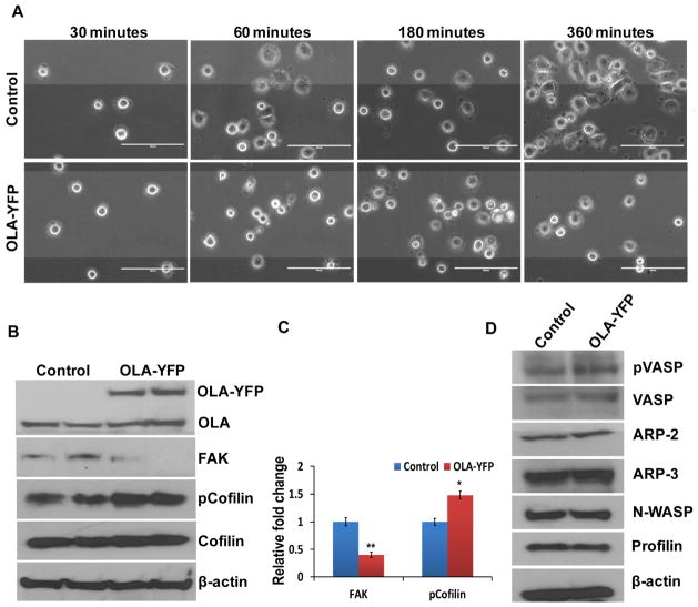
Figure 4. Overexpression of OLA1 results in delayed cell adhesion and spreading.
A, HeLa cells were transfected with an OLA1-expressing plasmid (OLA-YFP) or the YFP-expressing (control) vector for 48 hours, and re-plated on fibronectin-coated plates for cell adhesion assay. Cell images were taken at 30, 60, 180, and 360 minutes post-plating. Scale bar indicates 100 μm. B, Immunoblot analysis of the HeLa cells transfected with OLA1-YFP and the control vectors. The ectopic expression of OLA1 was confirmed by the presence of a higher molecular weight OLA1-YFP band on top of the endogenous OLA1 band. The same blot was re-probed for total FAK, total cofilin, and Ser3 phosphorylation of cofilin levels. β-actin was used as a loading control.. C, Densitomeric analysis of immunoblots shows fold changes of the FAK and pCofilin levels in OLA1 deficient cells compared to control cells. Data represent mean ± SD values from 3 independent experiments. **, p < 0.001; *, p < 0.05. D, Additional immunoblot analysis of the transfected HeLa cells for cytoskeletal signaling proteins as indicated. These data represent at least three independent experiments.

