Fig. 4.
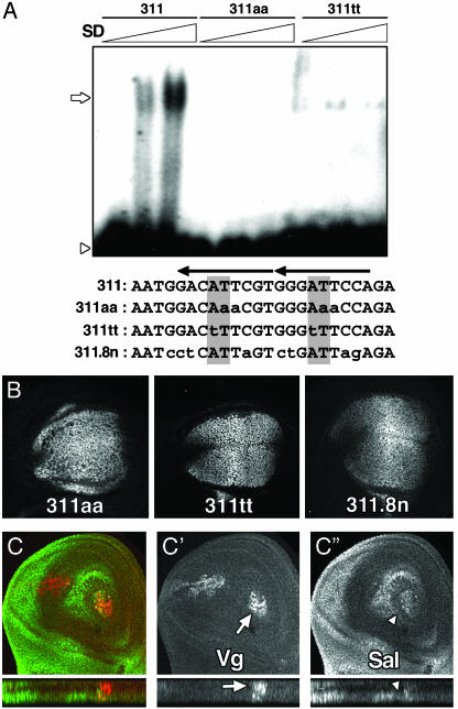
Sd–Vg complex is not essential for sal expression in the wing blade. (A) EMSA radiograms of bacterially expressed Sd protein. Wild-type oligonucleotide 311 shifts in the presence of increasing quantities of Sd. Binding is abolished by mutation 311aa or greatly diminished by mutation 311tt. Below the panel, the above oligonucleotide sequences are shown, as well as the sequence of the 311.8n mutant. The mutations are indicated in lowercase. The AT core from the TEA domain (TEF1, TEC1, ABAA) consensus sequences is shaded, and arrows indicate the Sd consensus binding sites. Open arrows indicate Sd binding, whereas open arrowheads indicate the free probe. (B) β-gal expression driven by mutant salE/Pv in third-instar imaginal discs. Sal expression in the same discs is not shown. (C) Expression of Sal (green) and Vg (red) in early third-instar wing imaginal discs with large territories mutant for vg. Red (C′) and green (C″) channels are shown in black and white. Despite the absence of Vg, Sal is expressed in the blade. Orthogonal sections below C′ show the remaining Vg-expressing cells (arrow). Around them, sal-expressing cells appear in a different plane (C″, arrowhead).

