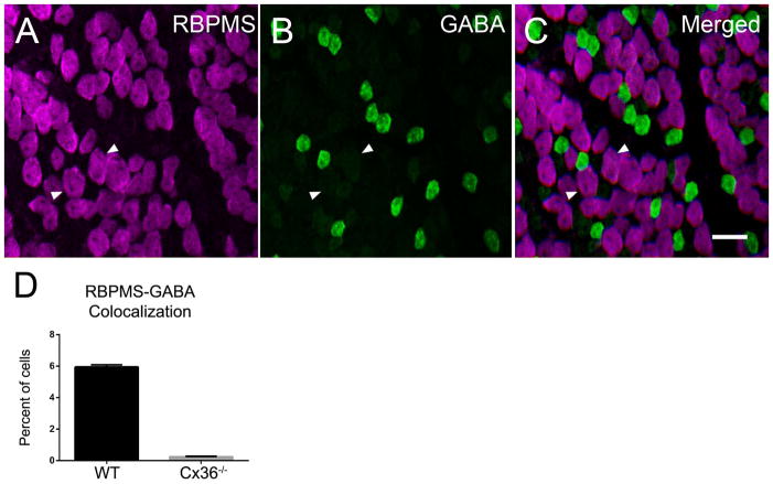
Figure 11.
The majority of RBPMS cells in the Cx36−/− mouse retina did not contain GABA immunoreactivity (A–D), although there were a few RBPMS cells with weak GABA immunoreactivity. Arrowheads indicate cells containing both RBPMS and weak GABA immunoreactivity. A: RBPMS. B: GABA. C: Merged image. D: Percentage of cells expressing both RBPMS and GABA immunoreactivity in wild-type (WT) and Cx36−/− whole-mounted retinas. Three wild-type (WT) and Cx36−/− retinas were evaluated. Plane of focus in GCL. z-step = 0.65 μm. 9 optical sections were compressed for viewing. RBPMS antibody GP15029. Scale bar = 20 μm.

