Figure 12.
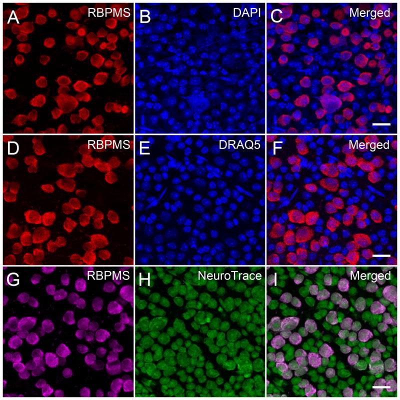
RBPMS immunoreactivity was mainly expressed in medium and large somata in the mouse GCL. Mouse whole-mounted retinas immunostained with RBPMS antibodies and counterstained with DAPI, DRAQ5 or NeuroTrace. DAPI and DRAQ5 labeled all nuclei, and NeuroTrace labeled RGC and displaced amacrine cell somata in the GCL. A, D, G: RBPMS. B: DAPI. E: DRAQ5. H: NeuroTrace. C, F, I: Merged images. Plane of focus in GCL for all images. z-step = 0.97 μm. 8 optical sections were compressed for viewing. RBPMS antibody GP15029. Scale bar = 20 μm.

