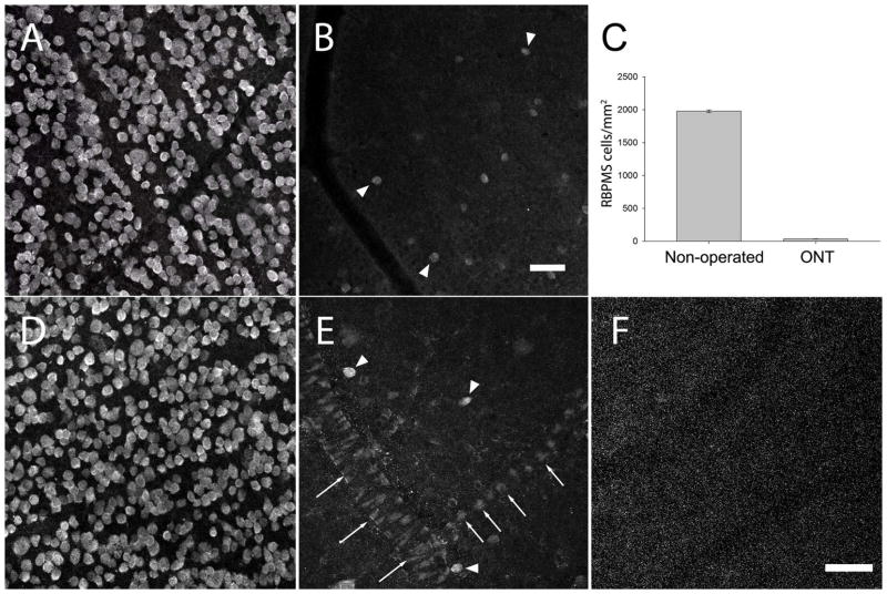
Figure 16.
RBPMS-immunoreactive cells in the GCL of the rat retina following unilateral optic nerve transection (ONT). A: GCL of the contralateral, non-operated retina. B: GCL of an axotomized retina 21 days after ONT (arrowheads indicate RBPMS immunoreactive cells). C: RBPMS cell density in the GCL decreased ~98% compared to the non-operated retina. RBPMS cell density was determined in 3 non-operated and 3 ONT eyes. D: GCL of the non-operated retina used as the control for the study illustrated in E and F. E: GCL of an axotomized retina 21 days after ONT (arrowheads indicate RBPMS cells). Note weak RBPMS immunostaining by endothelial cells of the blood vessels. F: RBPMS antibody preabsorbed with RBPMS immunizing peptide blocks immunolabeling of all cells including endothelial cells in the ONT retina. z-step = 1.1 μm. 12–14 optical sections were compressed for viewing. RBPMS antibody GP15029. Scale bar = 50 μm.

